
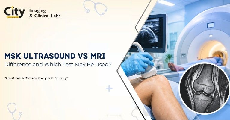
MSK Ultrasound vs MRI: What Is the Difference and Which Test May Be Used?

- Key Takeaways
- What Is an MSK Ultrasound Scan?
- What Is an MRI Scan?
- MSK Ultrasound vs MRI: Key Differences
- When May an MSK Ultrasound Be Used?
- When May an MRI Be Used?
- Which Test Is Better: MSK Ultrasound or MRI?
- Can MSK Ultrasound and MRI Be Used Together?
- MSK Ultrasound for Sports and Soft-Tissue Injuries
- Is MSK Ultrasound Safe?
- Is MRI Safe?
- MSK Ultrasound Scan in Delhi
- Frequently Asked Questions
- Final Takeaway
Musculoskeletal (MSK) imaging helps doctors evaluate problems involving muscles, tendons, ligaments, joints, soft tissues and other structures around the bones. Two commonly used imaging tests are MSK ultrasound and MRI. Although both can provide detailed information about musculoskeletal conditions, they work differently and are useful for different clinical situations.
MSK ultrasound uses high-frequency sound waves to create real-time images of muscles, tendons, ligaments and other soft tissues, while MRI uses a strong magnetic field and radio waves to produce highly detailed images of soft tissues, joints, cartilage, bone marrow and deeper structures. The choice between the two depends on the body part being examined, symptoms, suspected condition and the information the doctor needs.
If you are looking for an MSK Ultrasound Scan in Delhi, the appropriate test should generally be selected based on your doctor's clinical assessment and the specific area being evaluated.
Key Takeaways
- MSK ultrasound uses sound waves and shows muscles, tendons and ligaments in real time, even while you move the joint.
- MRI uses a magnetic field and radio waves and shows deeper structures, cartilage and bone marrow in fine detail.
- Neither test uses ionizing radiation.
- They are complementary; one is not always better than the other.
- Your doctor chooses the test based on the body part, symptoms and suspected condition.
What Is an MSK Ultrasound Scan?
An MSK ultrasound scan, or musculoskeletal ultrasound, is a non-invasive imaging test used to examine muscles, tendons, ligaments, joints and other soft tissues.
During the examination, a radiologist or trained healthcare professional places a handheld ultrasound probe on the skin over the area being examined. The probe sends high-frequency sound waves into the body and receives returning echoes, which are converted into images on a monitor.
One important advantage of MSK ultrasound is that it can provide real-time imaging. The examiner can observe certain structures while the patient moves the affected joint or muscle. This can be useful when evaluating some tendon problems, joint movement and soft-tissue abnormalities.
MSK ultrasound may be used to assess areas such as:
- Shoulder
- Elbow
- Wrist and hand
- Hip
- Knee
- Ankle and foot
- Muscles and tendons
- Certain superficial soft-tissue abnormalities
What Is an MRI Scan?
MRI, or Magnetic Resonance Imaging, uses a powerful magnetic field and radiofrequency energy to create detailed images of structures inside the body.
MRI can provide excellent visualization of many soft tissues and deeper structures. Depending on the body part and clinical question, MRI may be used to assess muscles, tendons, ligaments, cartilage, joints, nerves, bone marrow and other tissues.
Unlike ultrasound, MRI does not use sound waves to produce images.
MRI may be particularly useful when the suspected problem involves structures that are deep inside the body or when a more comprehensive assessment of a joint or surrounding tissues is required. You can see our MRI scan facilities in Delhi for the types of MRI available.
MSK Ultrasound vs MRI: Key Differences
The main difference between MSK ultrasound and MRI is the technology used and the type of information each examination can provide.
| Feature | MSK Ultrasound | MRI |
|---|---|---|
| Imaging technology | High-frequency sound waves | Magnetic field and radio waves |
| Radiation | No ionizing radiation | No ionizing radiation |
| Imaging | Real-time imaging | Detailed cross-sectional images |
| Movement assessment | Can be performed dynamically | Usually performed in a fixed position |
| Superficial tendons | Very useful | Very useful |
| Deep structures | More limited | Excellent visualization |
| Examination time | Usually shorter | Usually longer |
| Accessibility | Generally widely available | Availability may be more limited |
| Claustrophobia | Not usually an issue | Can be an issue for some patients |
| Contrast | Usually not required | Sometimes required depending on the examination |
These differences do not mean that one test is always better than the other. They are complementary imaging techniques, and the appropriate test depends on the clinical situation.
When May an MSK Ultrasound Be Used?
An MSK ultrasound may be considered when the doctor needs to evaluate a superficial soft-tissue structure or wants to observe a structure dynamically.
For example, ultrasound may be useful for assessing certain tendon abnormalities, muscle injuries, superficial masses and some joint-related conditions.
It can also allow the examiner to compare the affected side with the opposite side during the examination.
Another benefit is real-time assessment. For certain conditions, the patient may be asked to move the affected area while the radiologist observes the relevant structures.
MSK ultrasound can also be useful in some image-guided procedures, where real-time imaging helps the healthcare professional identify the target area.
When May an MRI Be Used?
MRI may be considered when the doctor needs detailed information about deep soft tissues, joints, cartilage, bone marrow, ligaments or other structures that may not be adequately assessed with ultrasound.
For example, an MRI may be requested for certain suspected:
- Ligament injuries
- Cartilage abnormalities
- Meniscus problems
- Deep muscle injuries
- Joint disorders
- Bone marrow abnormalities
- Complex soft-tissue conditions
MRI can also provide a broader view of the anatomy surrounding a joint or affected region, such as an MRI of the knee, shoulder, hip or elbow.
For patients with persistent symptoms where the cause is unclear, the doctor may determine that MRI is appropriate after considering the examination findings and previous imaging results.
Which Test Is Better: MSK Ultrasound or MRI?
There is no single test that is universally better. MSK ultrasound and MRI have different strengths, and the appropriate examination depends on the medical question.
For example, ultrasound may be particularly useful for a superficial tendon that needs dynamic assessment. MRI may provide more comprehensive information when the suspected abnormality involves deep structures, cartilage, bone marrow or multiple tissues around a joint.
In some situations, a doctor may initially recommend ultrasound and consider MRI if additional information is needed. In other cases, MRI may be selected from the beginning based on the symptoms and suspected diagnosis.
Therefore, patients should not select between MSK ultrasound and MRI based only on price or convenience. The imaging test should match the suspected condition and the information required by the treating doctor.
Can MSK Ultrasound and MRI Be Used Together?
Yes. In some clinical situations, ultrasound and MRI can complement each other.
For example, an ultrasound examination may provide a quick assessment of a superficial tendon and allow dynamic evaluation. If the doctor needs additional information about deeper structures or the overall joint, MRI may subsequently be recommended.
The two examinations provide different types of information, so having one test does not necessarily make the other unnecessary.
MSK Ultrasound for Sports and Soft-Tissue Injuries
MSK ultrasound can be useful in the evaluation of certain sports-related and soft-tissue injuries.
Athletes and physically active individuals may experience problems involving muscles, tendons and other soft tissues. Depending on the location and suspected injury, ultrasound can provide real-time information about the affected structure.
For example, an ultrasound examination may be used to evaluate certain tendon injuries around the shoulder, elbow, knee or ankle.
However, more complex injuries may require MRI when deeper structures, cartilage, ligaments or bone marrow need to be evaluated.
Is MSK Ultrasound Safe?
MSK ultrasound does not use ionizing radiation. It uses sound waves to produce images and is generally considered a safe imaging technique when performed appropriately.
Because the examination is external and does not normally require entering a scanner, it is also generally well tolerated.
However, whether ultrasound is appropriate depends on the clinical question. Your doctor or radiologist can determine whether it can provide the information required for your condition.
Is MRI Safe?
MRI does not use ionizing radiation. However, because MRI uses a strong magnetic field, patients need to inform the healthcare team about certain implants, medical devices, metal fragments or other relevant medical history before the examination.
Some MRI examinations may also involve contrast material. If contrast is recommended, the healthcare team will assess whether it is appropriate for the patient.
Patients should always follow the preparation instructions provided by the diagnostic centre and healthcare professional.
MSK Ultrasound Scan in Delhi
If you are searching for an MSK Ultrasound Scan in Delhi, City Imaging provides diagnostic imaging services for patients who need musculoskeletal imaging.
The exact type of ultrasound examination depends on the body part being evaluated and the clinical indication. Depending on the symptoms, a doctor may recommend an ultrasound of a specific tendon, muscle, joint or soft-tissue region.
Before booking an examination, it is useful to confirm:
- The exact type of MSK ultrasound required
- The body part to be examined
- Whether any preparation is necessary
- The available appointment timings
- The current test price
- Whether a doctor's prescription or referral is required
You can also read our guides on MRI of the LS spine, MRI of the whole spine and browse the Ultrasound and MRI Scan categories.
Frequently Asked Questions
Is MSK ultrasound the same as MRI?
No. MSK ultrasound and MRI are different imaging tests. Ultrasound uses high-frequency sound waves, while MRI uses a magnetic field and radio waves. Both can be used to evaluate musculoskeletal problems but provide different types of information.
Which is better for tendon problems, ultrasound or MRI?
Both can be useful for tendon assessment. Ultrasound can provide real-time and dynamic evaluation of many superficial tendons, while MRI can provide detailed information about deeper structures and surrounding tissues. The appropriate test depends on the suspected condition.
Does MSK ultrasound use radiation?
No. MSK ultrasound does not use ionizing radiation. It uses high-frequency sound waves to create images.
Does MRI use radiation?
No. MRI does not use ionizing radiation. It creates images using a strong magnetic field and radiofrequency energy.
How long does an MSK ultrasound take?
The duration can vary depending on the body part being examined and the complexity of the examination. Your diagnostic centre can provide the expected examination time when you schedule the appointment.
Do I need an MRI after an MSK ultrasound?
Not necessarily. An MRI may be recommended only when additional information is needed or when the suspected condition is better evaluated with MRI. The decision is made based on the clinical findings and imaging requirements.
Is MSK ultrasound painful?
It is generally painless. You may feel slight pressure from the probe, especially over a tender area, and you may be asked to move the joint during the scan.
Do I need to prepare for an MSK ultrasound or MRI?
Preparation depends on the test. Ultrasound of a joint or tendon usually needs little preparation, while MRI may need you to remove metal objects and share your implant history. Follow the instructions given by your doctor or diagnostic centre.
Can I have an MRI if I have a pacemaker or metal implant?
It depends on the type of device or implant. Always tell the healthcare team before an MRI so that they can check whether it is safe for you.
Which test should I choose without a doctor's advice?
It is best not to choose by price or convenience alone. Your doctor will advise the test that matches your symptoms and the information needed.
Final Takeaway
MSK ultrasound and MRI are complementary imaging techniques rather than direct substitutes. MSK ultrasound is particularly useful for real-time evaluation of many superficial muscles, tendons and soft tissues, while MRI provides highly detailed images of deeper tissues, joints, cartilage, ligaments and bone marrow.
If your doctor has recommended an MSK Ultrasound Scan in Delhi, confirm the specific body part and examination required before booking. The right imaging test is the one that can provide the information needed to evaluate your symptoms accurately.






.jpeg)

.png)




Comments List